Welcome to Matrix Education
To ensure we are showing you the most relevant content, please select your location below.
Select a year to see courses
Select a year to see available courses
Need help exchanging biology knowledge for good marks? Well in this article, we walk you through the organisation of living things so you can make Band 6 results as easy as you do gas exchange (breathing)!
This module looks at how cells are organised into a hierarchical system to help organisms survive. We will look at the syllabus content NESA expects students to gain mastery of.
You will also learn about how nutrients and gases are transported in animals and plants.
Cells are arranged differently in unicellular, multicellular and colonial organisms.
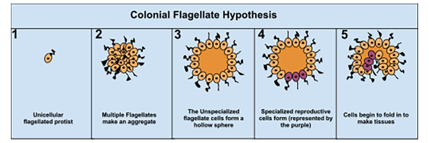
The Colonial Flagellate Hypothesis proposes that the cells of unicellular organisms aggregated to form a colony. The cells in the colony could survive on their own if separated. Over millions of years, some cells differentiated into different types and, eventually, they became dependent on each other. These were the first multicellular organisms.
As the complexity of multicellular organisms increased, the need for hierarchical organisation of cells became crucial for organism survival.
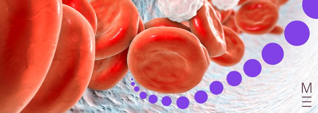
Different cell types in the body arise from undifferentiated cells known as stem cells. Under the control of gene expression, they develop structures specific to their role in the organism. Examples include red blood cells which have haemoglobin proteins and a biconcave shape to efficiently transport oxygen. Cells are grouped together to form tissues, layers of different types of tissue make up organs. Organs with similar functions are grouped into organ systems which make up an organism. Organ systems help deliver nutrients and remove wastes from the organism.
Autotrophs such as plants produce organic compounds from inorganic compounds obtained from the environment through photosynthesis. Heterotrophs such as animals use cellular respiration for energy production. They cannot produce organic compounds on their own and must consume other organisms to satisfy their nutritional requirements.
Plants have systems and structures such as root, stem, and leaves that help carry out functions such as gas exchange, water and nutrient absorption. Macroscopic structures of plants can be examined by dissecting plant material.
| Plant Structures | |
| Root | Root hairs are fine, hair-like structures that increase the surface area of the root in contact with soil to facilitate absorption. This includes water absorption through osmosis (via passive transport) and mineral absorption (via active transport).
|
| The root tip is constantly growing and consists of a meristematic and elongation region.The root tip is constantly growing and consists of a meristematic and elongation region. • Meristematic region – the site of cell division | |
| The root cap protects the growing tip of the root. | |
| Special root structures like nodules contain nitrogen-fixing bacteria that harness nitrogen from the air. | |
| Stem | Xylem tissue consists of dead cells that form a hollow tube which moves water and ions in a unidirectional way from roots to shoots. |
| Phloem tissue consists of living tissue that transports sugars in a bidirectional way from the leaves to the rest of the plant. | |
| Leaves | External structure of the leaf is made up of: • Waxy cuticle to reflect off heat and reduce water loss |
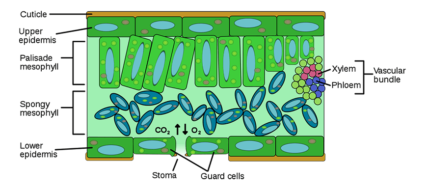
| Internal structures of the leaf include: • Upper and lower epidermises contain stomata (microscopic pores) that control gas exchange and water loss. | |
Plants lack specialised gas exchange systems and instead have structures that allow different parts of the plant to obtain gases from their immediate environment. These structures include stomata, lenticels, and roots.
Gas exchange in leaves is facilitated through stomata which are microscopic pores surrounded by guard cells that control when they open and close to reduce water loss from the plant. Carbon dioxide enters through the stomata and diffuses through spongy mesophyll cells into the palisade mesophyll where most photosynthesis occurs. Oxygen and water exit through the open stomata.
Animals have respiratory systems with structures that aid in gas exchange. These structures differ between animals, but some important structural features are always the same: moist and thin membranes, rich blood supply and a large surface area.
Insects, for example, have a network of holes called spiracles and branching tubes called tracheoles. The tracheoles increase the internal surface area of the insect to maximise diffusion rate of gases around their body. Diffusion is also facilitated by the insect’s abdominal movement that flushes air in and out of the tracheal system.
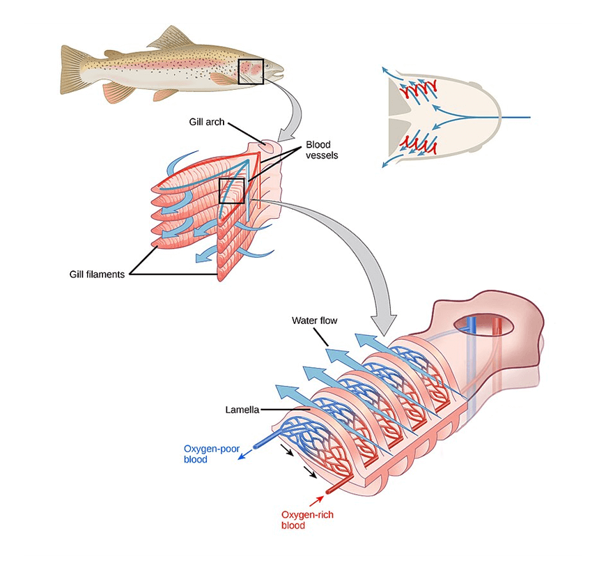
In fish, gas exchange is performed by the gills which contain gill arches to support filaments and lamellae. The lamellae increase the surface area available for gas exchange. Since the concentration of dissolved oxygen is lower in water compared to air, gills use a counter-current exchange mechanism to increase the efficiency of oxygen transfer. This mechanism maintains a constant concentration gradient by ensuring that the blood in fish always contains lower oxygen concentration than the water flowing over it, leading to increased efficiency of oxygen diffusion.
Amphibians such as frogs acquire various gas exchange structures depending on their development stage. Frogs have a larval stage (aquatic stage) and an adult stage (semi-terrestrial). During the larval stage, they develop internal and external gills which work the same way as the gills in fish for gas exchange. At the adult stage, simple lungs are developed instead. Simple lungs are not efficient, so frogs also exchange gases through the skin and within the mouth.
In mammals such as humans, the gas is exchanged through a respiratory system which contains structures such as lungs (alveoli) and airways (trachea, bronchi and bronchioles).
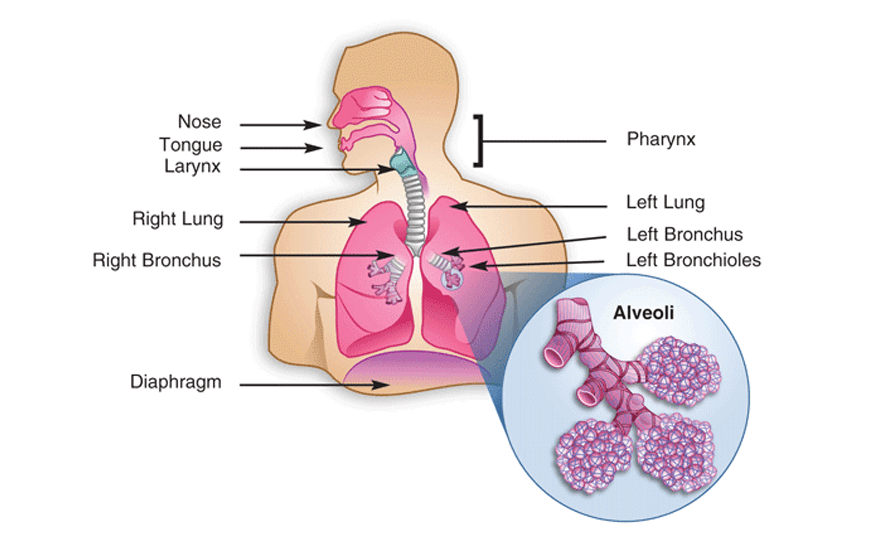
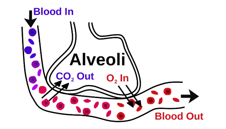
Air containing a mixture of oxygen and other gases is inhaled and then filtered through the trachea. The trachea branches into tubes called bronchi and bronchioles that lead into the lungs. The branching of these tubes increases surface area to maximise gas exchange. Once in the lungs, an exchange of oxygen and carbon dioxide takes place in the alveoli which are small sacs surrounded by blood vessels. Blood transports oxygen to body cells where it is used in cellular respiration to produce energy. Carbon dioxide is produced as a waste product and is transported by the blood to the alveoli to be exhaled.
During this term, you will also cover the different digestive systems and dentition (teeth) found in herbivores, omnivores, and carnivores. The function of the digestive system is to breakdown food, releasing essential nutrients such as sugars and proteins so they can be absorbed into the blood stream.
In humans, the digestive system is made up of the following:
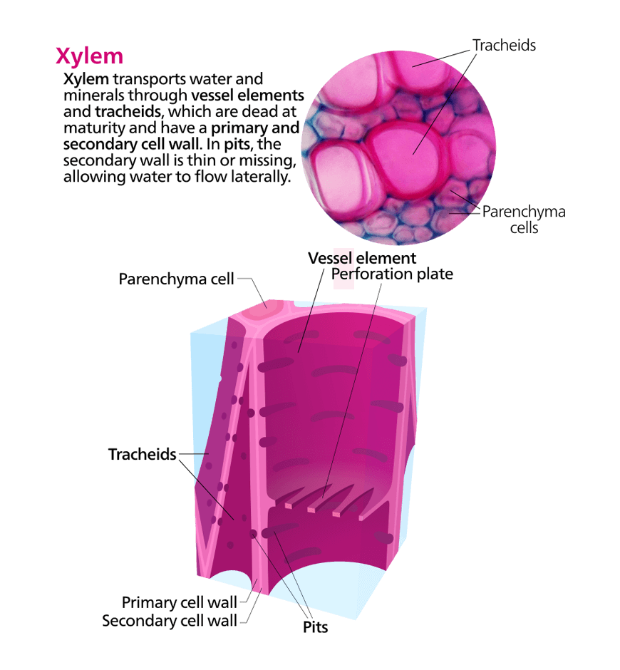
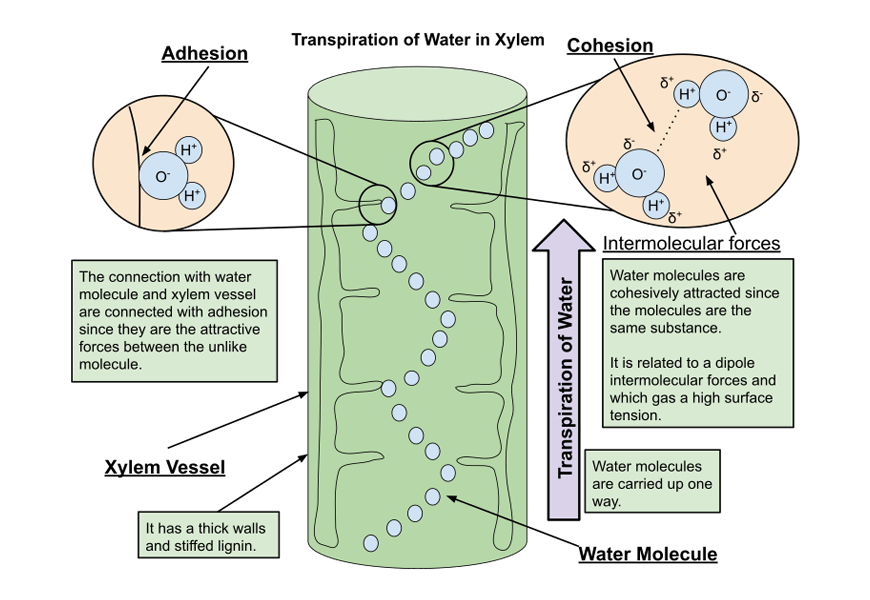
The transport of water and sugars in plants is carried out by vascular bundles which are made up of two main tissue types: xylem and phloem. The xylem tissue is made up of dead cells (tracheids) that lie end to end to form hollow tubes. The cell wall of the xylem is reinforced with lignin to provide more rigidity and prevent its collapse as water moves upwards through it. Xylem transports water and mineral ions only in a unidirectional way (i.e. from roots to leaves only).
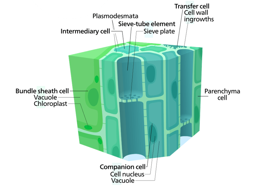
Sugars are distributed around the plant through phloem tissue in a bidirectional way. Phloem tissue is made up of living cells called sieve tubes. These cells have sieve plates to allow the flow of sugars throughout the phloem and companion cells that provide energy for the active transport of sugars in and out of the phloem.
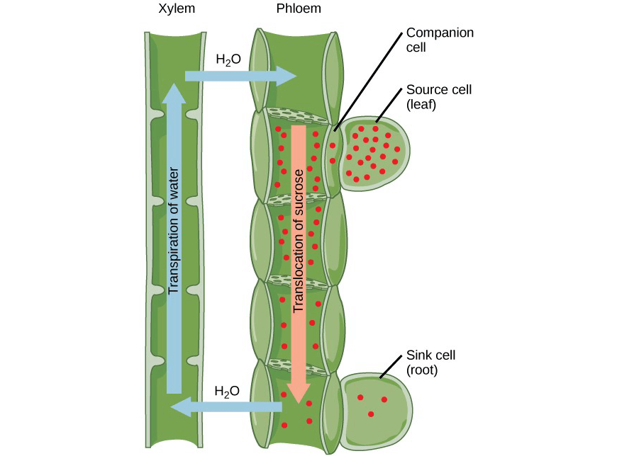
The Pressure-Flow hypothesis also known as source-to-sink theory explains how sugars move through the phloem tissue. The source is the site where sugars are made (i.e. leaves) and the sink is any part of the plant that require sugar for growth and other metabolic functions.
Sugar production through photosynthesis in plant leaves results in phloem loading due the high sugar concentration at the source. This causes water in the xylem to flow into the phloem by osmosis. The pressure of water movement from the source to the sink forces the sugars to move along with it. At the sink, sugar is unloaded to parts of plants that need it, causing water to move out of the phloem and into the xylem again by osmosis.
The circulatory system in animals is a network system that aids in the transport of gases and nutrients to nourish the cells while removing metabolic wastes such as urea. There are two types of circulatory systems: open and closed circulatory systems.
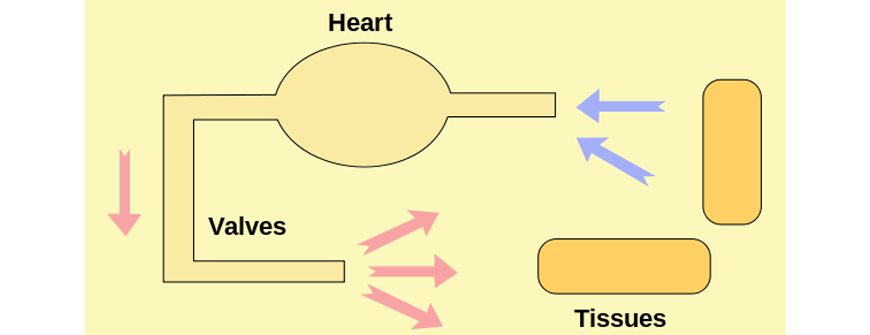
In an open circulatory system, open ended vessels fill a cavity known as haemocoel with blood-like fluid called haemolymph that is pumped by a simple tube heart. Exchange of nutrients and wastes occurs when cells and tissues interact with this haemolymph. The open system is commonly seen in invertebrates such as insects as shown below. Haemolymph does NOT however transport gases since invertebrates have a separate system for gas exchange.
Organisms that are larger in size such as mammals rely on a closed circulatory system as it is more efficient in transporting materials. In a closed circulatory system, the heart pumps blood containing gases, nutrients and wastes around the body through closed vessels. These vessels include arteries, veins, and capillaries.
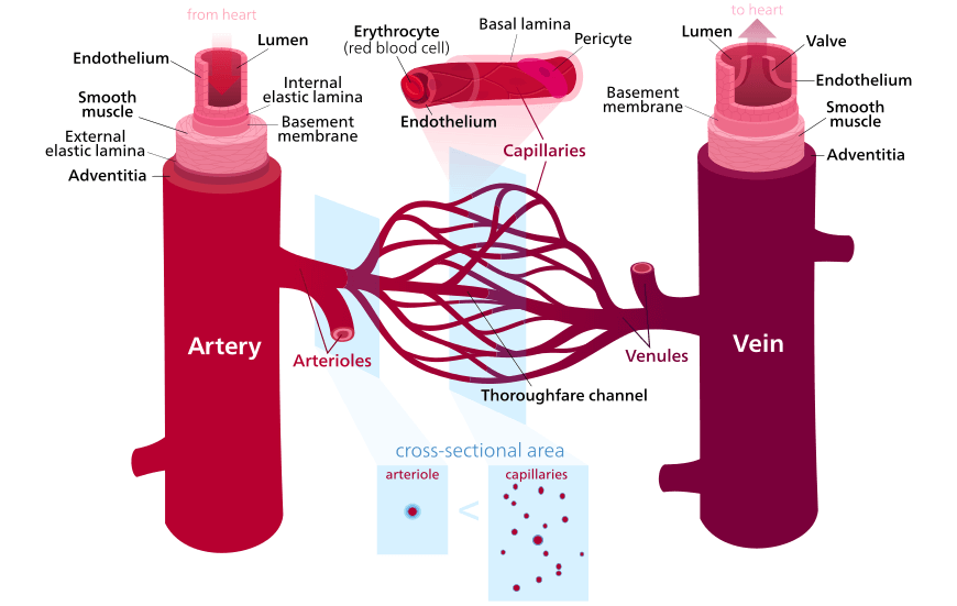
Unlike invertebrates, the heart of vertebrates is more efficient as it contains chambers. In humans, there are four chambers that separate oxygenated blood from de-oxygenated blood. This means that one system flows from the heart to lungs and back (Pulmonary circulation) and the other system flows from the heart to the body and back (Systemic circulation). This is described as a double circulatory system.
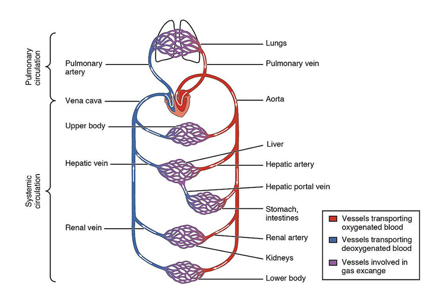
The composition of the blood changes as it moves around an organism to distribute nutrients, gases and wastes. In animals, as blood moves through the lungs, the concentration of oxygen increases while carbon dioxide decreases due to the gas exchange in the alveoli.
In plants, fluid composition in the xylem and phloem changes as it moves around the plant. This includes an increase in sugar concentration in leaves due to photosynthesis and a decrease in sugar concentration in any growing parts of the plants that uses sugar. You will cover more examples about changes in composition in further details in your class.
Structures in plants and animals can be visualised using different technologies such as the light microscope. In animals, the light microscope can produce micrographs to visualise blood components and cells. You will be required to draw scaled diagrams and perform calculations to estimate the size of the cells.
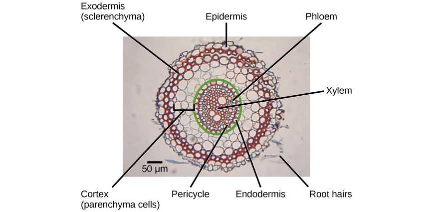
Microscopic structures in plants can be observed using a light microscope. An example of a transverse section of a root is shown below.
© Matrix Education and www.matrix.edu.au, 2025. Unauthorised use and/or duplication of this material without express and written permission from this site’s author and/or owner is strictly prohibited. Excerpts and links may be used, provided that full and clear credit is given to Matrix Education and www.matrix.edu.au with appropriate and specific direction to the original content.