Welcome to Matrix Education
To ensure we are showing you the most relevant content, please select your location below.
Select a year to see courses
Select a year to see available courses
Trying to figure out the building blocks of good biology marks? In this guide, we give you a breakdown of cells as the basis of life, the first module of Biology for Stage 6.
This module focuses on cells and cell types, their internal organelles and general functions. We will look at the syllabus content NESA expects students to gain mastery of.
In order to understand cells, you are going to learn about what distinguishes cells from one another and how cells coordinate their activities within their internal and external environment.
Cells are significant as they are the very basis of life. In 1839, scientists Theodor Schwann and Matthias Schleiden developed the tenets of classical cell theory, stating that:
There are two types of cells: Prokaryote and Eukaryote.


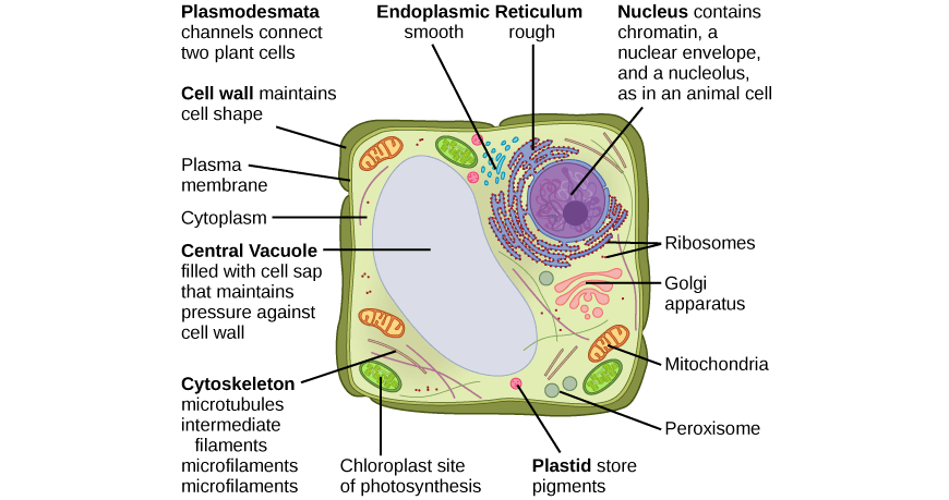
Plant specific structures include a cell wall, central vacuole and chloroplasts which are not found in an animal cell.
Eukaryotes can be unicellular or multicellular organisms, whereas, prokaryotes are always unicellular. The following characteristics further help us differentiate between Eukaryotic and Prokaryotic cells.
| Characteristic | Prokaryote | Eukaryote |
| DNA storage | Free-floating in the cytoplasm Long circular nucleoid and smaller plasmids | Located in the nucleus. Composed of multiple chromosomes |
| Membrane-bound organelles | None | Many e.g. nucleus, Golgi apparatus, endoplasmic reticulum. |
| Cell Wall | Present in all bacteria and archaea. | Found only in plants, fungi and some protists. |
| Size | 0.1-0.5 µm | 10-100 µm |
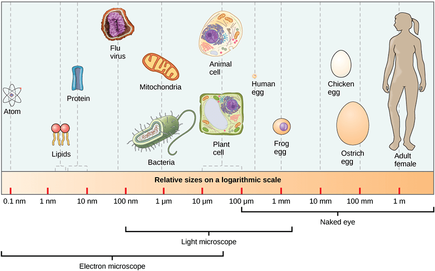
Since cell organelles are too tiny to be seen by the naked eye (as shown on the scale below), different technology such as microscopes, staining techniques, and lighting techniques have enabled us to visualise cells and their contents.
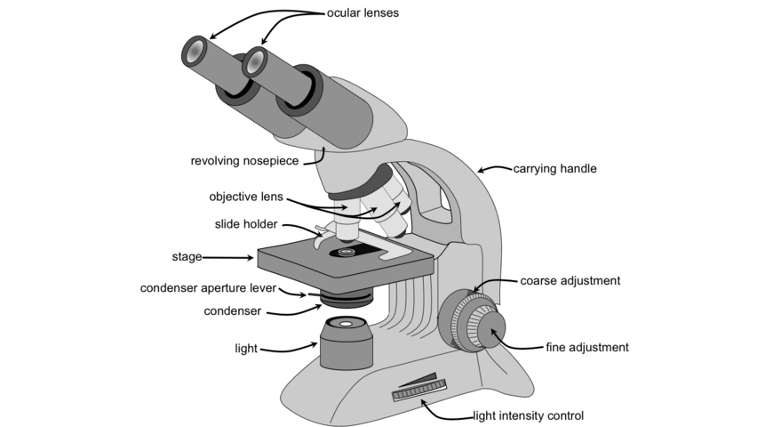
The first type of microscope invented was the light microscope which uses light to visualise cells ranging in size from 1mm to 100nm. It contains magnifying glasses inside the ocular lens and objective lens that provide magnification and resolution for improved cell visualisation.
The light microscope has limitations in terms of image detail. Therefore, an electron microscope can be used instead to resolve objects less than 1 nm and produces increased resolution and magnification of up to 2,000,000X! The electron microscope uses electron beams instead of light and can come in two types:
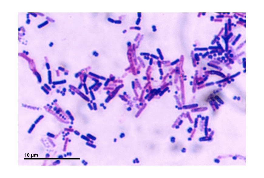
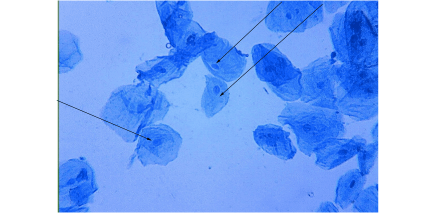
Staining techniques such as dyes allows for the staining of different cellular structures, for example:
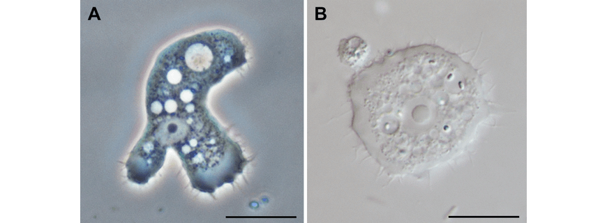
Lighting techniques such as phase-contrast microscopy also allows scientists to gain improved image quality of cells. Phase-contrast enhances the contrast to allow visualisation of cell contents and thus can be advantageous for observation of biological processes. The following comparison is of an amoeba under phase contrast (A) versus bright field microscopy (B).
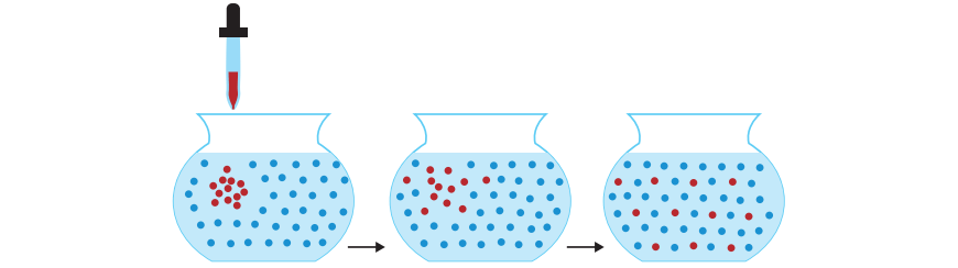
Diffusion is an important concept in biology. Diffusion is the habit of liquids and gases to move from an area of high concentration to an area of low concentration. Osmosis is the diffusion of water across a semi-permeable membrane. Diffusion and osmosis help to explain how nutrients and wastes are transported in and out of cells.
Cells must obtain nutrients to survive while expelling waste materials that can become toxic if accumulated in large amounts. This is facilitated by the cell membrane that aids in exchanging these materials within their internal and external environment. The membrane is composed of phospholipid bilayer containing millions of phospholipid molecules with hydrophilic heads facing outwards towards water and hydrophobic tails facing inwards away from the water.
Embedded in the membrane are structures of lipids, carbohydrates and proteins, as shown below.
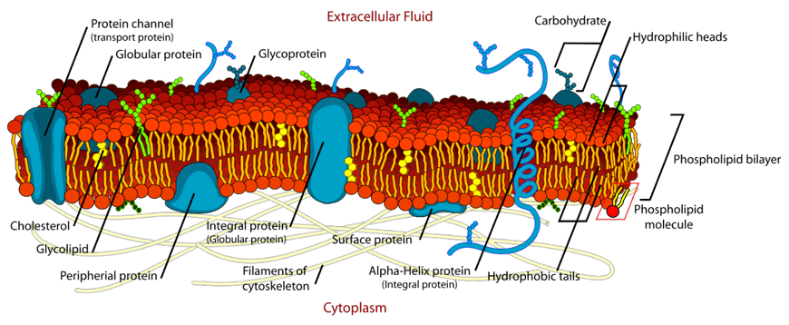
Some small, non-polar, and neutral substances including water, oxygen and carbon dioxide can easily move through the membrane via passive transport. Passive transport does NOT require energy in the form of ATP. Passive transport moves water molecules through osmosis and ions through facilitated diffusion using protein channels embedded within the cell membrane. These substances move down their concentration gradient from high concentration to low concentration areas. To gain a better understanding of this, you will investigate conducting an experiment to observe and model diffusion and osmosis.
On the other hand, large, polar, and charged molecules such as lipids and carbohydrates cannot easily cross the cell membrane and therefore move via active transport which requires both the protein channels and ATP. Active transport moves substances up their concentration gradient from areas of low to high concentration. In order to infect a cell, viruses must trick the cell into letting them through the cell membrane!
At times, cells require movement of large amounts of materials into the cells using a process called endocytosis. In endocytosis, the plasma protein folds around the extracellular material to form a vesicle that engulfs the material into the inside of the cell.
The three types of endocytosis include:
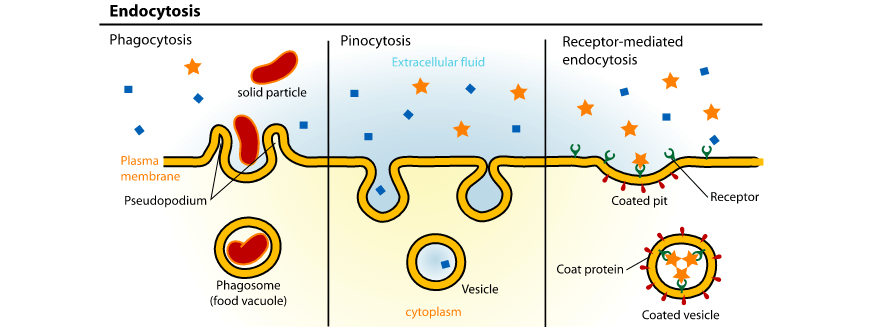
Exocytosis, on the other hand, exports materials such as hormones and waste products outside of the cell. This process is the reverse of endocytosis where the materials are expelled in vesicles that fuse with the membrane to be transported out of the cell.
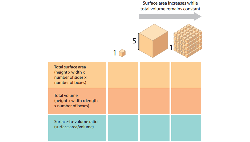
Because they rely on diffusion and osmosis, the exchange of materials into and out of the cell is limited by the size of the cell. Cells are generally smaller than 1mm in size and if they were to grow beyond that, metabolic wastes would accumulate and nutrients would be inefficiently transported. As a result, cells must maintain a high Surface Area to Volume ratio (SA:V).
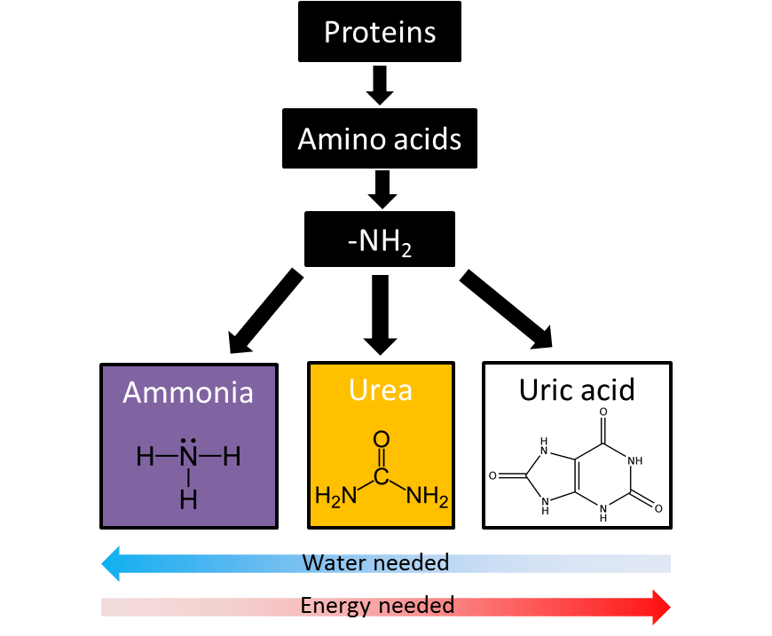
Metabolic wastes such as nitrogenous wastes must be removed from the organism’s body for normal cellular functioning. The breakdown of proteins in the cell can produce nitrogenous wastes such as ammonia which is highly toxic and requires high water levels for its excretion. Nitrogenous wastes are thus converted into different forms depending on the availability of water for improved excretion rate. For example, aquatic species have an abundant supply of water and thus excrete this waste in the form of ammonia, whereas, in mammals, the nitrogenous waste must be converted into urea as it is less toxic and requires less water for its excretion.
Plants are autotrophs that obtain nutrients by performing photosynthesis within their chloroplasts. The numerous flattened discs in the chloroplast increases the surface area thus allowing higher rate of photosynthesis to occur.
Animals are typically heterotrophs that consume other organisms to obtain nutrients. They then produce chemical energy (ATP) through cellular respiration that occurs in the mitochondria. The inner mitochondrial membrane is folded to increase its surface area and thus maximise cellular respiration rate.
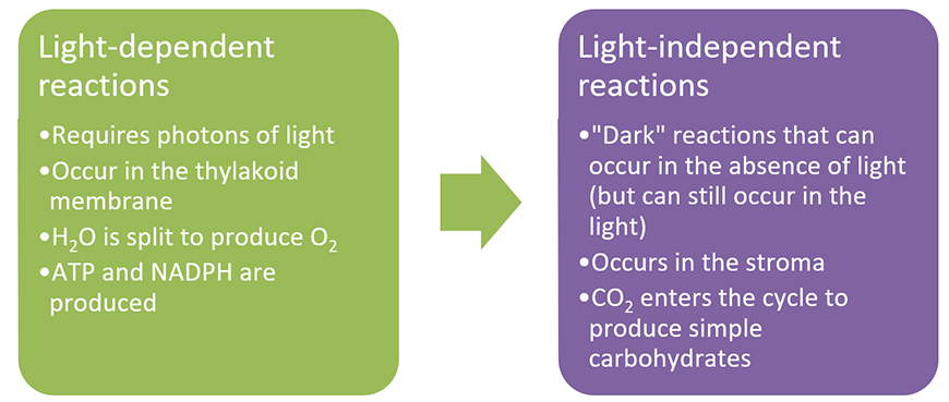
As mentioned, plants are autotrophs that use photosynthesis to produce their own food. There are two processes involved in photosynthesis: the light-dependent stage and the light-independent stage. The summary of these stages is as follows:
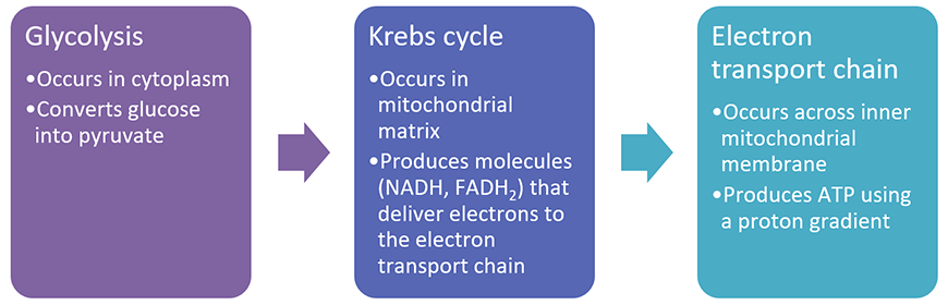
Animals are heterotrophs that use the mitochondria for cellular respiration to generate ATP from glucose (plants can also undergo cellular respiration!). There are three processes involved in cellular respiration: Glycolysis, the Krebs Cycle, and the Electron Transport Chain. The summary of these stages is as follows and will be covered in more details during the class.
Cells respond to energy demand through the process of metabolism. Metabolism is the collection of life-sustaining chemical reactions which involves anabolism and catabolism. Catabolism breaks down complex nutrients such as glucose in order to release energy for cellular use, while, anabolism is the synthesis of simple nutrients into more complex ones for energy storage.
In order to speed metabolic reactions, cells have enzymes which are a type of protein that catalyse reactions by lowering the energy level required to start the reaction.

There are two models of how enzyme reactions work: the “lock and key” model where the active site of the enzyme matches the substrates perfectly (as above) and the “induced fit” model where the active site changes shape slightly to fit the substrate.
Since enzymes are proteins, they possess a specific three-dimensional shape that must be maintained for them to function efficiently. Factors such as temperature, substrate concentration, and pH level all have an impact on enzyme activity. At high temperatures or at high/low pH, enzymes denature – they change shape and can no longer function.
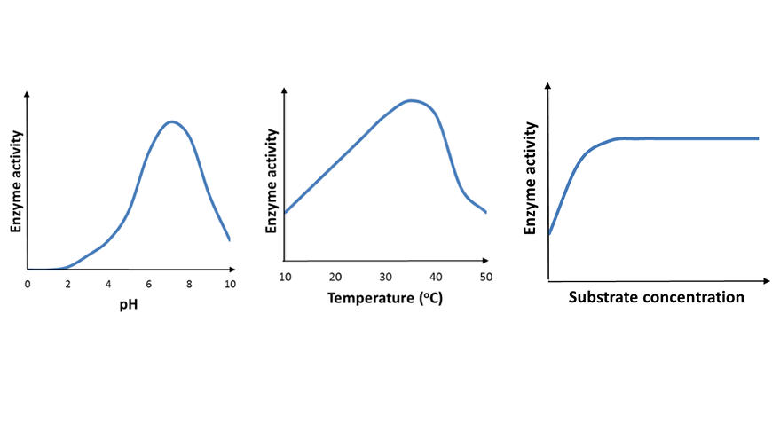
During your class, you will carry out a first-hand investigation to better understand how enzyme activity is impacted by the environment of the cell.
© Matrix Education and www.matrix.edu.au, 2025. Unauthorised use and/or duplication of this material without express and written permission from this site’s author and/or owner is strictly prohibited. Excerpts and links may be used, provided that full and clear credit is given to Matrix Education and www.matrix.edu.au with appropriate and specific direction to the original content.